Saline may also be transferred using a bacteriological loop. On the opposite side, a small drop of sterile saline is taken. Marking the slide makes it convenient to identify the surface of the slide that contains the smear.
#Gram positive vs gram negative cells on a slide free
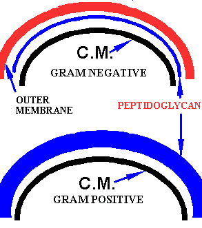
This theory is probably not widely accepted.Īnother theory such as presence of Magnesium ribonucleate in Gram positive bacteria and its absence in Gram negative bacteria has not received widespread acceptance.Īt the end of Gram staining, Gram positive bacteria appear violet or purple whereas Gram negative bacteria appear pink.Ī grease free glass slide is taken and a circle is marked one side of the slide using a wax pencil or a glass marker pen. Slightly lower cytoplasmic pH (2.0) in Gram positive bacteria helps the basic dye to bind more efficiently than the Gram negative bacteria who have slightly elevated pH (3.0). On the other hand, alcohol dehydrates the Gram positive bacteria closing the pores and shrinking the cell wall and thus prevents outflow of dye-iodine complex. Typically, gram positive bacteria have thicker cell walls (approximately 40 layers of peptidoglycan) compared to Gram negative bacteria, which have only 1-2 layers of peptidoglycan.Īlcohol or acetone, which is used as a decolorizer dissolves lipids in the membranes since Gram negative bacteria have an additional membrane (apart from cytoplasmic membrane), more lipids are lost forming pores through which the dye-iodine complex escapes during decolorization. Various theories have been put forth to explain the differences among bacteria that are exploited by this staining technique some are more acceptable than others. Those bacteria that resist decolorization and retain the primary dye are called Gram positive and those bacteria that gets decolorized and then get counterstained are called Gram negative. When stained with a primary stain and fixed by a mordant, some bacteria are able to retain the primary while others get decolorized by a decolorizer.

There are four components of Gram stain primary stain, mordant, decolorizer, and counterstain. The original formulation comprised of aniline gentian violet, Lugol's iodine, absolute alcohol and Bismarck brown. Gram stain was described by Danish bacteriologist Hans Christian Gram in 1884 to differentiate Streptococcus pneumoniae from Klebsiella pneumoniae in lung tissue.

Different bacteria stain differently to a common staining procedure. Such staining methods are called differential staining methods, these include Gram staining and acid fast staining. Some staining techniques utilize these differences to stain the bacteria differently. Bacteria vary in certain physiological and biological properties.


 0 kommentar(er)
